Every time you nod, turn to check your blind spot, or look down at a screen, tiny deep muscles and tough connective tissues at the top of your neck coordinate that movement. Scientists have been mapping this region in detail and have discovered that it is a closely connected web where muscles, ligaments and the protective covering of the spinal cord all interact. Understanding this hidden system helps explain why the way we use our necks – and the positions we keep them in – can sometimes play a part in how our head and neck feel day to day.
What is the myodural bridge?
A 2020 cadaver study by Sillevis and Hogg examined the region just under the back of the skull to explore how these tissues might relate to neck pain and certain headaches. Your brain and spinal cord are wrapped in three membranes called meninges, with the “dura” mater forming the tough outer layer. This layer lines the inside of the skull and continues down the spinal canal, acting like a protective inner jacket that helps contain the fluid around the brain and spinal cord. Because it contains many nerve endings, the dura can be sensitive to stretch, pressure and irritation.
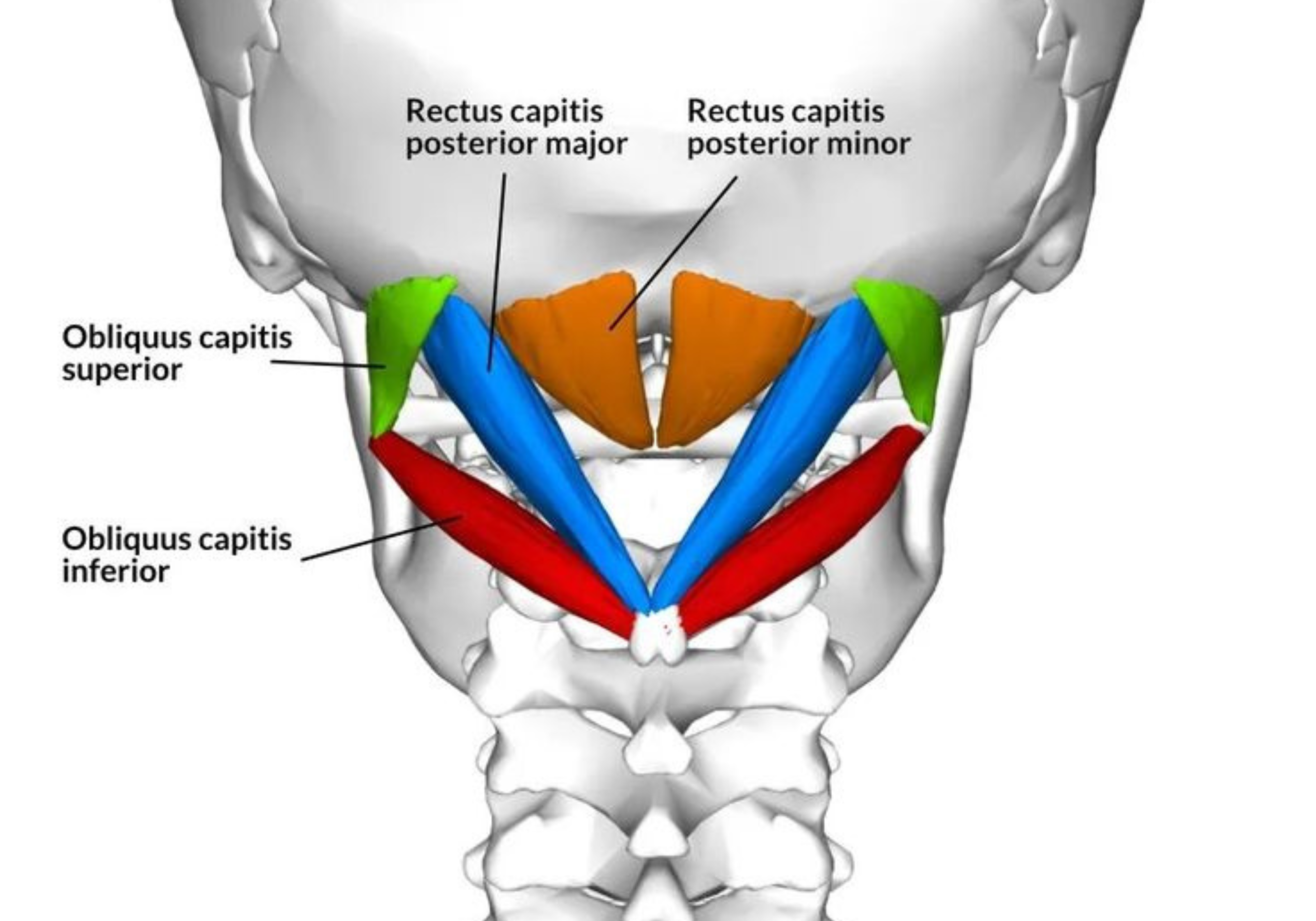
The 2020 study focused on the upper cervical spine where the head meets the neck: the occiput (base of the skull), the atlas (C1) and the axis (C2). The researchers examined the small suboccipital muscles and the nuchal ligament, which contribute to fine control of head position and support the back of the neck. They identified a continuous network of connective tissue linking muscles, ligaments, bones and the dura. In particular, they described a broad, tough sheet of tissue extending from the suboccipital muscles towards the back of the spinal canal at C1, blending with the tissue surrounding the dura and forming what is known as a myodural bridge – a structural bridge between muscle and dura.
How the upper neck talks to your brain?
A review by Enix, Scali and Pontell (2014) brings together the available anatomical studies, imaging work and small clinical observations on this “cervical myodural bridge” and asks what this might mean in day‑to‑day life. The authors describe the myodural bridge as an inherent link between the deepsuboccipital muscles and the dura. These deep muscles are rich in proprioceptive sensors that inform your brain about the precise position and movement of your head in space. The review proposes that when these muscles activate, they transmit a measured, functionally helpful tension through the myodural bridge, which may assist in keeping the dura and spinal cord stable while allowing them to glide smoothly during normal movements. The authors also suggest that disruption of this system – for example through injury, persistent overactivity or changes in muscle properties – could play a role in some neck‑related headaches and dizziness, though this remains a hypothesis rather than a proven cause–effect relationship.

What the research really shows (and what it doesn’t)?
Both papers have important limitations that are clearly acknowledged. The 2020 study is based on cadaveric tissue, so it can describe structure and possible movement but cannot determine how these relationships behave in living people with pain. The 2014 article is a narrative review that integrates anatomy, imaging and small clinical reports, but it is not a large clinical trial and does not directly test treatments, exercises or everyday habits. Crucially, neither paper measures smartphone use, laptop posture, or working from the sofa or bed, so they cannot state that these behaviours directly cause headaches via the myodural bridge; instead, they offer a plausible anatomical and proprioceptive framework that clinicians may consider alongside broader biopsychosocial factors when assessing neck‑related symptoms.
Clinical perspective on neck posture and headaches
My clinical opinion, grounded in what these two papers actually show, is this: the upper neck seems to have a clever system where deep muscles “talk” directly to the dura via the myodural bridge and help keep the spinal cord stable as you move. Persistent or abnormal loading of these muscles (after an injury, or possibly from long periods in non‑neutral postures such as staring down at a phone or slumping with a laptop) may, over time, interfere with that system. For some people, that could be one factor in neck‑related headaches and balance or coordination issues.
These papers do not prove that scrolling on a phone or working from bed directly causes headaches, but they do offer a coherent mechanical story for why caring for the upper neck is clinically relevant. In practice, that means paying attention to how this region moves, how long it is held in one position, and how effectively the deep muscles are working to support and guide that movement. Alongside broader lifestyle and psychosocial factors, regularly moving and gently loading this area is a reasonable, low-risk way to support its proprioceptive and mechanical function.

Practical ways to care for your upper neck
One simple option is to include a comfortable suboccipital stretch in your routine. Lying on your back with your head supported, imagine making a subtle “nodding” motion as if saying “yes” to a very small question, gently tucking the chin without forcing it. You should feel a mild lengthening at the base of the skull rather than a big pull through the front of the neck; hold this light stretch for 10–20 seconds, breathe quietly, then relax, repeating a few times as tolerated.
As always, anyone with significant pain, dizziness, neurological symptoms or medical concerns should seek individual assessment before starting new exercises
References
Enix DE, Scali F, Pontell ME. “The cervical myodural bridge, a review of literature and clinical implications.” Journal of the Canadian Chiropractic Association, 2014;58(2):184‑192
Sillevis R, Hogg R. Anatomy and clinical relevance of sub occipital soft tissue connections with the dura mater in the upper cervical spine. PeerJ. 2020 Aug 10;8:e9716